New Discovery Shows How Gamma Delta T Cells Can Destroy Cancer
6 September 2023
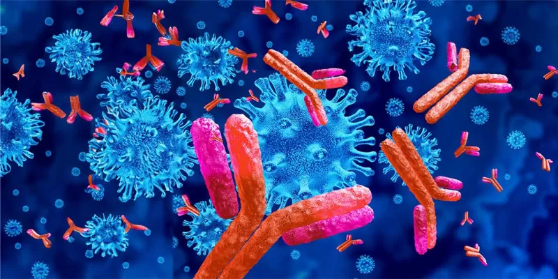
The groundbreaking research from Gladstone Institutes and UC San Francisco, using CRISPR technology, found that Gamma delta T cells, target a molecular complex called butyrophilin on cancer cells. Cancer cells, due to hyperactive cholesterol production, make this complex more accessible, marking them for destruction by gamma delta T cells.
Gamma delta T cells, specific components of our immune system, have a natural knack for detecting and destroying cancer cells. Intriguingly, cancer patients with elevated levels of these cells often have a more optimistic prognosis. This discovery, published in Nature, offers promising avenues for future immunotherapies.
T cells play a vital role in our body's defenses, continually scouring for threats from harmful bacteria to mutated cancer cells. They identify these threats through a protein called the T cell receptor. A particular group of T cells, known as gamma delta T cells, have shown exceptional prowess in identifying cancer cells across the body. But how they performed this feat at a molecular level remained a mystery. Now researchers have determined the conditions that enable these T cells to recognize cancer cells.
"We used the power of CRISPR genome editing to get fundamental insights into what can make cancer cells recognizable to gamma delta T cells so they can be targeted for elimination. Our work opens the door to thinking about how this pathway could eventually be targeted in future immunotherapies," says Dr. Alex Marson, PhD, director of the Gladstone-UCSF Institute of Genomic Immunology and senior author of the study.
Through rigorous experimentation, the team discovered that these gamma delta T cells identified cancer cells by targeting a molecular complex named butyrophilin. This complex appears on many human cells, both healthy and otherwise. What makes cancer cells different, they found, is hyperactive cholesterol production, which subsequently activates the butyrophilin complex, marking them for detection by gamma delta T cells.
Furthermore, the research unveiled that stress and diminished energy production in cancer cells increase the presence of butyrophilin on their surfaces. Treating tumor cells with drugs that emulate stress amplifies this effect, rendering them more susceptible to gamma delta T cell detection and subsequent destruction.
"We knew that gamma delta T cells recognize their target cells in a very different way from conventional T cells, but the field has had some trouble figuring out exactly how the gamma delta T cells were recognizing the cancer cells," says Murad Mamedov, PhD, postdoctoral scholar at Gladstone and first author of the study.
Mamedov, Marson, and their collaborators utilized CRISPR technology to disrupt thousands of genes in lymphoma cells, systematically testing which gene disruptions impacted the ability of gamma delta T cells to kill cancer cells. They verified that the gamma delta T cells recognized a complex of molecules named butyrophilins,which were previously shown to be targeted by gamma delta T cells. Researchers believes that this revelation will pave the way for enhancing the efficacy of cancer immunotherapies.
Erin J. Adams, PhD, another collaborator, added, "This research demystifies the remarkable capabilities of gamma delta T cells, some of the immune system's most lethal operatives." The study's findings, published in the esteemed journal Nature, not only present fresh insights into the workings of gamma delta T cells but also open the door to novel therapeutic strategies that capitalize on their cancer-fighting prowess.
Abstract of the research
CRISPR screens decode cancer cell pathways that trigger γδ T cell detection
Abstract: γδ T cells are potent anticancer effectors with the potential to target tumours broadly, independent of patient-specific neoantigens or human leukocyte antigen background1,2,3,4,5. γδ T cells can sense conserved cell stress signals prevalent in transformed cells2,3, although the mechanisms behind the targeting of stressed target cells remain poorly characterized. Vγ9Vδ2 T cells—the most abundant subset of human γδ T cells4—recognize a protein complex containing butyrophilin 2A1 (BTN2A1) and BTN3A1 (refs. 6,7,8), a widely expressed cell surface protein that is activated by phosphoantigens abundantly produced by tumour cells. Here we combined genome-wide CRISPR screens in target cancer cells to identify pathways that regulate γδ T cell killing and BTN3A cell surface expression. The screens showed previously unappreciated multilayered regulation of BTN3A abundance on the cell surface and triggering of γδ T cells through transcription, post-translational modifications and membrane trafficking. In addition, diverse genetic perturbations and inhibitors disrupting metabolic pathways in the cancer cells, particularly ATP-producing processes, were found to alter BTN3A levels. This induction of both BTN3A and BTN2A1 during metabolic crises is dependent on AMP-activated protein kinase (AMPK). Finally, small-molecule activation of AMPK in a cell line model and in patient-derived tumour organoids led to increased expression of the BTN2A1–BTN3A complex and increased Vγ9Vδ2 T cell receptor-mediated killing. This AMPK-dependent mechanism of metabolic stress-induced ligand upregulation deepens our understanding of γδ T cell stress surveillance and suggests new avenues available to enhance γδ T cell anticancer activity.


Comments
No Comments Yet!