AI-Supported Skin Scanning Revolutionizes Diabetes Diagnosis and Monitoring

Digital Health |
12 December 2023
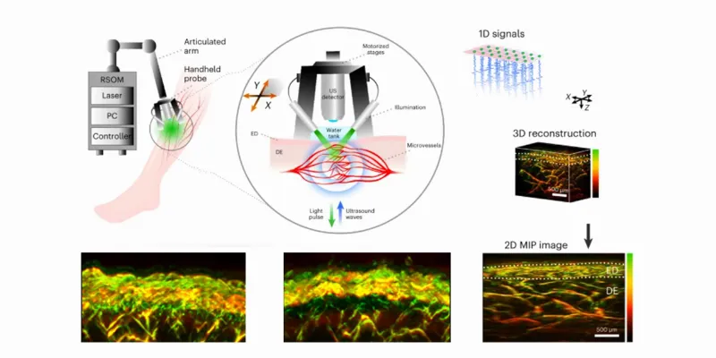
In a groundbreaking study, researchers developed a new, faster, non-invasive method using AI-supported skin scanning for diabetes diagnosis. They utilized RSOM mesoscopy combined with AI to analyze microvascular changes in the skin of patients. This accurate method, providing detailed images, marks a significant advancement in diagnosing and monitoring the severity of diabetes, potentially transforming patient care.
Researchers at the Technical University of Munich (TUM) and Helmholtz Munich have made a significant breakthrough in the assessment of diabetes severity. Utilizing a novel imaging method called Raster-Scan Optoacoustic Mesoscopy (RSOM), combined with the precision of Artificial Intelligence (AI), they can now analyze the microvascular changes in the skin, a common consequence of diabetes. This innovative approach promises to greatly improve the management and treatment of diabetes.
The Study's Findings
In a study involving 75 diabetic patients and a control group, RSOM was used to capture images of leg blood vessels. AI algorithms then identified 32 significant changes in the skin's microvasculature, such as variations in vessel branches and diameter. These changes are crucial indicators, providing essential information about the disease's progression and severity.
The Science Behind RSOM
Optoacoustic imaging is a cutting-edge technique in which light pulses create ultrasound waves within tissue. These waves, generated by the expansion and contraction of tissue around light-absorbing molecules such as hemoglobin, are then converted into detailed images of blood vessels. The study, led by Professor Vasilis Ntziachristos and his team at TUM and Helmholtz Munich, demonstrates that RSOM, unlike other non-invasive methods, achieves unparalleled depth and detail.
Towards a New Era of Diabetes Monitoring
"With RSOM, we can now quantitatively describe the effects of diabetes," says Professor Ntziachristos. "With the emerging ability to make RSOM portable and cost effective, these findings open up a new way for continuous monitoring of the status of those affected - more than 400 million people worldwide. In the future, with fast and painless examinations, it would take just a few minutes to determine whether therapies are having an effect, even at home environments."
The Advantages of RSOM Over Traditional Methods
Traditional biopsy methods, while informative, are invasive and can distort the real condition of blood vessels. RSOM, on the other hand, offers a quick, non-invasive alternative that does not require radiation or contrast agents. It provides comprehensive data on different skin layers in a single, rapid measurement. This method revealed, for the first time, that diabetes impacts vessels in various skin layers differently, a crucial insight for understanding the disease.
The research delves deeper into the technical aspects, discussing the segmentation of skin layers and microvasculature using machine learning. It highlights the sensitivity of dermal layer features to various stages of diabetes. The 'microangiopathy score,' derived from the 32 most relevant features, accurately predicts the presence of diabetes with a high degree of precision, underscoring the potential of RSOM in discovering new diabetes biomarkers and monitoring the condition.
The collaboration of advanced optoacoustic imaging and AI in assessing diabetes marks a significant stride in medical science. RSOM stands as a beacon of hope, offering a non-invasive, accurate, and quick method to understand and manage diabetes, a disease that affects millions worldwide.
Abstract of the research
Dermal features derived from optoacoustic tomograms via machine learning correlate microangiopathy phenotypes with diabetes stage
Abstract: Skin microangiopathy has been associated with diabetes. Here we show that skin-microangiopathy phenotypes in humans can be correlated with diabetes stage via morphophysiological cutaneous features extracted from raster-scan optoacoustic mesoscopy (RSOM) images of skin on the leg. We obtained 199 RSOM images from 115 participants (40 healthy and 75 with diabetes), and used machine learning to segment skin layers and microvasculature to identify clinically explainable features pertaining to different depths and scales of detail that provided the highest predictive power. Features in the dermal layer at the scale of detail of 0.1–1 mm (such as the number of junction-to-junction branches) were highly sensitive to diabetes stage. A ‘microangiopathy score’ compiling the 32 most-relevant features predicted the presence of diabetes with an area under the receiver operating characteristic curve of 0.84. The analysis of morphophysiological cutaneous features via RSOM may allow for the discovery of diabetes biomarkers in the skin and for the monitoring of diabetes status.







Comments
No Comments Yet!