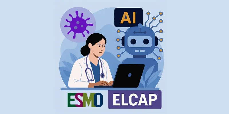
Artificial Intelligence Detects Pancreatic Cancer More Accurately Than Radiologists

An international PANORAMA study shows that artificial intelligence (AI) can outperform radiologists in detecting pancreatic cancer on routine CT scans. Across 3,440 patients, AI achieved an AUROC of 0.92 compared with 0.88 for 68 radiologists, detecting more tumors while generating fewer false positive results. These findings underscore AI’s potential to support earlier diagnosis of pancreatic ductal adenocarcinoma using standard-of-care imaging.
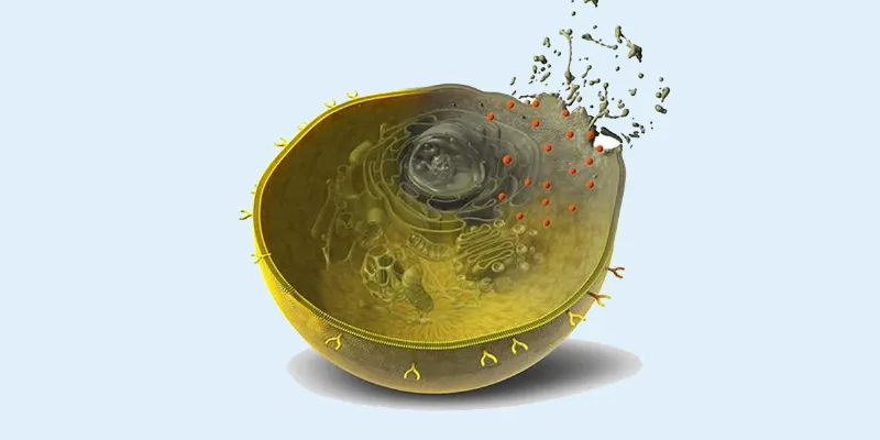
Pancreatic ductal adenocarcinoma remains one of the most lethal solid tumors in clinical practice. Fewer than ten percent of patients survive beyond five years, largely because the disease is usually diagnosed too late for curative surgery. Contrast enhanced computed tomography is the cornerstone of diagnosis, yet early stage tumors are notoriously difficult to identify.
“Our findings show that AI can be a valuable support for radiologists in the challenging task of detecting pancreatic cancer,” says Dawid Rutkowski, PhD student at the Department of Clinical Science, Intervention and Technology. “We now aim to move forward with prospective studies where the AI system is tested in clinical practice, but with great caution and care.”
Building a global benchmark for AI
That gap is addressed by PANORAMA, an international, paired, confirmatory observational study published in The Lancet Oncology. Led by investigators from Radboud University Medical Center, Karolinska Institutet, and the University of Bergen, the project set out to create a robust, open source benchmark for pancreatic cancer detection on standard of care CT scans.
The scope of the effort is notable. In total, 3,440 patients were included, drawn from tertiary care centers in Europe and the United States. More than 430 AI developers from 46 countries submitted algorithms to the benchmark. Rather than selecting a single model, the researchers combined the three best performing algorithms into a final AI system, aiming to represent the upper bound of current technical performance.
The system was trained and tuned on 2,310 patients and then evaluated on a fully sequestered testing cohort of 1,130 patients from centers in the Netherlands, Sweden, and Norway. A multi reader, multi case observer study compared the AI directly with 68 radiologists from 40 institutions across 12 countries, with a median of nine years of clinical experience. The reference standard relied on histopathology and at least three years of clinical follow up, strengthening the validity of the findings.
AI edges past human expertise
The primary endpoint was diagnostic accuracy, measured by the area under the receiver operating characteristic curve. In the sequestered testing cohort, the AI system achieved an AUROC of 0.92. In the subset used for the reader study, AI again reached an AUROC of 0.92, while the pooled performance of radiologists was 0.88.
Statistically, the AI system was both non inferior and superior to radiologists. Clinically, the difference translated into the detection of more tumors with fewer false positive findings. In a disease where missing an early lesion can mean the difference between resection and palliation, this margin matters.
From detection to surveillance
Beyond initial diagnosis, the researchers see potential applications in longitudinal care. Patients with pancreatic cysts or precursor lesions are often monitored with repeated imaging over years, a process that is resource intensive and vulnerable to subtle progression being missed.
Integrating AI into radiology workflows raises questions about generalizability, accountability, and overreliance on automated outputs. Prospective validation in real world settings will be essential before clinical adoption. The PANORAMA study does not claim to have solved pancreatic cancer. Still, the findings offer a credible glimpse of how AI could shift the diagnostic timeline, identifying tumors when surgery is still possible and survival curves can still bend.
Conducted with support from the European Union and without known conflicts of interest, and involving partners such as Cleveland Clinic, Mayo Clinic, Johns Hopkins University, and Massachusetts General Hospital, the study sets a new benchmark for how AI should be evaluated in oncology imaging.


Comments
No Comments Yet!